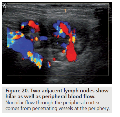
Breast Cancer Lymph Node Color Doppler Ultrasound Color Meanings

Normal Lymph Node On Ultrasound Lymph Nodes Typically Are Smooth Download Scientific Diagram
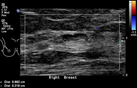
Color Doppler Sonography Characterizing Breast Lesions

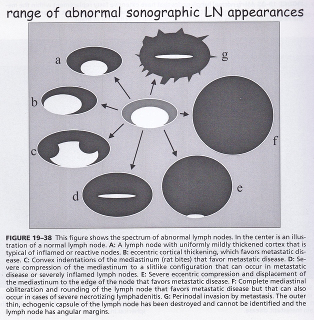
Typical Us Appearance Of Lymph Nodes Typical Reactive Node Image A Download Scientific Diagram
Reactive Vs Malignant Lymph Nodes Ultrasound Features Radiology Reference Article Radiopaedia Org

Pdf Ultrasonography Of Superficial Lymph Nodes Benign Vs Malignant
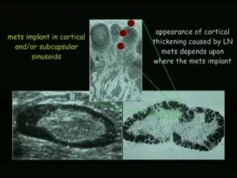
Ultrasound Evaluation Of Regional Lymph Nodes As An Extension Of The Breast Ultrasound Exam Youtube
B d three dimensional multiplanar reformatted images also show loss of the fatty hilum.
Breast cancer lymph node color doppler ultrasound color meanings.
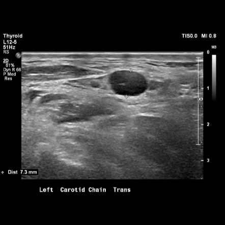
Breast Cancer Topic Lymph Nodes On Ultrasound
Intramammary Lymph Nodes Radiology Reference Article Radiopaedia Org

Lymph Node Matting Multiple Malignant Nodes Are Fused In A Single Ill Download Scientific Diagram

Nodal Echogenic Hilum A Benign Node With Echogenic Central Hilum B Download Scientific Diagram
Source : pinterest.com
